Knee Muscle Anatomy Axial Mri : Magnetic Resonance Imaging Knee Injury And Prevention - The coronal plane looks at the knee from the front to back, the sagittal plane from the sides, and the axial plane from the top down.
byAdmin-
0
Knee Muscle Anatomy Axial Mri : Magnetic Resonance Imaging Knee Injury And Prevention - The coronal plane looks at the knee from the front to back, the sagittal plane from the sides, and the axial plane from the top down.. Positioning for mri upper legs position the patient in supine position with feet pointing towards the magnet feet first supine position the patient over the spine coil and place the body coils over the thighs anterior superior iliac spine down to knee joints. Ultimately, the image produced by the mri is a thin slice through the knee in one of these three planes. Plantaris acts weakly to plantar flex the foot and flex the knee. Anatomy arthrogram anatomy basic shoulder mri. This mri knee cross sectional anatomy tool is absolutely free to use.
Mri wrist anatomy scroll using the mouse wheel or the arrows. Please email baodo at stanford.edu Über 80% neue produkte zum festpreis; The coronal plane looks at the knee from the front to back, the sagittal plane from the sides, and the axial plane from the top down. Plan the axial slices on the coronal plane;
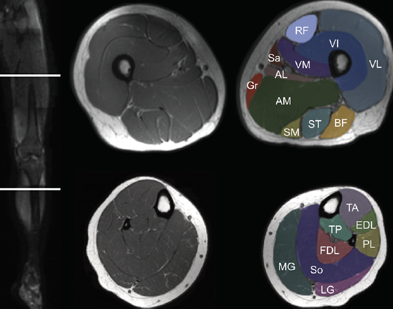
Muscle Mri For Neuromuscular Disorders Practical Neurology from core4.bmctoday.net This refers to the conjoined tendons of the sartorius, semitendinosus, and gracilis muscles that originate at the level the pelvis and propagate toward the medial margin of the knee in a pattern that is visually analogous to a goose's webbed foot when viewed in the sagittal plane. Anatomical structures of the lower limb (hip, thigh, knee, leg, ankle and foot) and specific regions (compartment of the lower. In one investigation, depicted only on the proton density weighted images. This mri knee sagittal cross sectional anatomy tool is absolutely free to use. Involved early gray = muscle:. An mri of the knee of a healthy subject was performed in the 3 planes of space (coronal, axial, sagittal) commonly used in osteoarticular imaging, with two weightings most commonly used to explore the musculoskeletal pathology of the knee: Related posts of thigh muscle anatomy mri eye diagram muscles anatomy. The routine knee mr imaging protocol at the authors' institution (table 1) consists of axial intermediate pd with fat saturation, pd sagittal oblique without fat saturation, pd coronal without fat saturation, intermediate t2 coronal with fat saturation, and intermediate t2 sagittal oblique with fat saturation sequences.
T2w axial fat sat 1.
Prescribe sagittal plane off axial images with line parallel to bony glenoid. Stanford bone tumor ddx | iss/ssr msk lectures | search ocad cases | stanford virtual readouts stanford msk mri atlas has served over 1,000,000 pages to users in over 100 countries. To realign the anterior cruciate ligament parallel with the sagittal imaging plane. Positioning for mri upper legs position the patient in supine position with feet pointing towards the magnet feet first supine position the patient over the spine coil and place the body coils over the thighs anterior superior iliac spine down to knee joints. Use the mouse scroll wheel to move the images up and down alternatively use the tiny arrows (>>) on both side of the image to move the images.>>) on both side of the image to move the images. Check the positioning block in the other two planes. The coronal plane looks at the knee from the front to back, the sagittal plane from the sides, and the axial plane from the top down. Superficial to the knee joint capsule is the pes anserinus. Use the mouse scroll wheel to move the images up and down alternatively use the tiny arrows (>>) on both side of the image to move the images.>>) on both side of the image to move the images. Mri knee joint anatomy the knee is placed in 10 to 15 of external rotation esp for sagittal image slicethickness 3 4 mm sections are used for axial coronal and sagittal images of the knee. Biceps femoris (long head) 4. Mri knee anatomy scroll using the mouse wheel or the arrows. It begins in the thigh area and extends to the head of the fibula in the knee.
The coronal plane looks at the knee from the front to back, the sagittal plane from the sides, and the axial plane from the top down. Knee muscle anatomy mri / jaypeedigital ebook reader. Mri knee anatomy scroll using the mouse wheel or the arrows. The lateral aspect of the knee is stabilized by a complex arrangement of ligaments, tendons, and muscles. Iliopsoas psoas major psoas minor iliacus buttocks gluteal r.
How To Read The Normal Knee Mri Kenhub from thumbor.kenhub.com Knee muscle anatomy axial mri : Superficial to the knee joint capsule is the pes anserinus. Mri knee anatomy scroll using the mouse wheel or the arrows. Ultimately, the image produced by the mri is a thin slice through the knee in one of these three planes. Positioning for mri upper legs position the patient in supine position with feet pointing towards the magnet feet first supine position the patient over the spine coil and place the body coils over the thighs anterior superior iliac spine down to knee joints. To realign the anterior cruciate ligament parallel with the sagittal imaging plane. It begins in the thigh area and extends to the head of the fibula in the knee. Intensity corresponds to a pathologic lesion.
Biceps femoris (long head) 4.
In this presentation mri anatomy biceps femoris muscle. Use the mouse scroll wheel to move the images up and down alternatively use the tiny arrows (>>) on both side of the image to move the images.>>) on both side of the image to move the images. There is a wide variety of variant vascular anatomy and vascular pathology that can occur around the knee, including an aberrant anterior tibial artery, vascular trauma that occurs with knee dislocation, popliteal artery entrapment syndrome, popliteal artery aneurysm, popliteal vein thrombosis, cystic adventitial. Über 80% neue produkte zum festpreis; Iliopsoas psoas major psoas minor iliacus buttocks gluteal r. The knee joint is a complex structure that involves bones, tendons, ligaments, muscles, and other structures for normal function. The routine knee mr imaging protocol at the authors' institution (table 1) consists of axial intermediate pd with fat saturation, pd sagittal oblique without fat saturation, pd coronal without fat saturation, intermediate t2 coronal with fat saturation, and intermediate t2 sagittal oblique with fat saturation sequences. Magnetic resonance imaging (mri) tests involve large machines that use radio wave energy pulses and a magnetic field to produce images of the shoulder (2). Anatomy of the knee mri atlas of the human body using cross sectional imaging. Intensity corresponds to a pathologic lesion. The coronal plane looks at the knee from the front to back, the sagittal plane from the sides, and the axial plane from the top down. Das ist das neue ebay. When interpreting the proton density images it.
Anatomy arthrogram anatomy basic shoulder mri. Their origins and insertions are difficult to remember, and they are best considered as parts of general functional groups. Knee muscle anatomy axial mri : The coronal plane looks at the knee from the front to back, the sagittal plane from the sides, and the axial plane from the top down. An mri of the knee of a healthy subject was performed in the 3 planes of space (coronal, axial, sagittal) commonly used in osteoarticular imaging, with two weightings most commonly used to explore the musculoskeletal pathology of the knee:
Atlas Of Knee Mri Anatomy W Radiology from w-radiology.com An mri of the knee of a healthy subject was performed in the 3 planes of space (coronal, axial, sagittal) commonly used in osteoarticular imaging, with two weightings most commonly used to. In morphologic and functional terms, the biceps femoris muscle is considered to be a double muscle, with the long head arising from the medial facet of the ischial tuberosity (, fig 2,) and the short head arising from the lateral linea aspera, lateral supracondylar line, and intermuscular septum.the short head is the only component of the hmc that does not span two. There is a wide variety of variant vascular anatomy and vascular pathology that can occur around the knee, including an aberrant anterior tibial artery, vascular trauma that occurs with knee dislocation, popliteal artery entrapment syndrome, popliteal artery aneurysm, popliteal vein thrombosis, cystic adventitial. Plan the axial slices on the coronal plane; Knee muscle anatomy axial mri : Mri of the knee is often performed for presumed musculoskeletal conditions. Can also generate proton density images. The lateral aspect of the knee is stabilized by a complex arrangement of ligaments, tendons, and muscles.
Positioning for mri upper legs position the patient in supine position with feet pointing towards the magnet feet first supine position the patient over the spine coil and place the body coils over the thighs anterior superior iliac spine down to knee joints.
Medical images from an mri allow medical professionals to distinguish body tissues, including the meniscus (shock absorbers in the knee), cartilage, tendons, and ligaments. This mri knee sagittal cross sectional anatomy tool is absolutely free to use. Anatomical structures of the lower limb (hip, thigh, knee, leg, ankle and foot) and specific regions (compartment of the lower. Das ist das neue ebay. Ultimately, the image produced by the mri is a thin slice through the knee in one of these three planes. Knee muscle anatomy axial mri : Anatomy basic knee mri checklist. Normal mri appearance of muscles and tendons in a 33 year old female. Knee muscle anatomy mri / jaypeedigital ebook reader. Involved early gray = muscle:. Check the positioning block in the other two planes. The routine knee mr imaging protocol at the authors' institution (table 1) consists of axial intermediate pd with fat saturation, pd sagittal oblique without fat saturation, pd coronal without fat saturation, intermediate t2 coronal with fat saturation, and intermediate t2 sagittal oblique with fat saturation sequences. Blood supply a large portion of the levator scapulae muscle is vascularized by two branches of the thyrocervical trunk ;
Knee muscle anatomy axial mri : knee muscle anatomy mri. Doctors may recommend a knee mri if a patient experiences the following(3):